. 2020 May 15;40(4):1107–1124. doi: 10.1148/rg.2020190145

Figure 14a.

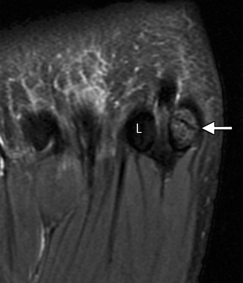
Bipartite sesamoiditis. (a–c) Axial (a) and sagittal T2-weighted FS (b) and sagittal (c) PDW images show medial sesamoiditis in a 24-year-old woman with pain at the plantar aspect of the right first MTPJ. There is a bifid appearance of the medial sesamoid with marrow edema (solid arrow in a and b) at both components. Smooth corticated margins and the presence of a waist at the superior margin of the sesamoid (arrow in c) are more suggestive of bipartite sesamoiditis rather than a fracture. Partial tearing of the medial SPL is also seen (dotted arrow). Radiographs were not available in this patient but are useful for assessment and follow-up. L = lateral sesamoid. (d, e) Dorsoplantar (d) and lateral (e) radiographs in a 15-year-old patient with plantar trauma show separation of two components at the medial sesamoid (dashed circles). This was a challenging case, with the proximal rounded margins of the distal component (arrowhead) suggestive of diastasis of a bipartite sesamoid rather than a fracture. Despite the challenges in differentiating the two conditions, fracture and diastasis are treated similarly with initial conservative management.

Bipartite sesamoiditis. (a–c) Axial (a) and sagittal T2-weighted FS (b) and sagittal (c) PDW images show medial sesamoiditis in a 24-year-old woman with pain at the plantar aspect of the right first MTPJ. There is a bifid appearance of the medial sesamoid with marrow edema (solid arrow in a and b) at both components. Smooth corticated margins and the presence of a waist at the superior margin of the sesamoid (arrow in c) are more suggestive of bipartite sesamoiditis rather than a fracture. Partial tearing of the medial SPL is also seen (dotted arrow). Radiographs were not available in this patient but are useful for assessment and follow-up. L = lateral sesamoid. (d, e) Dorsoplantar (d) and lateral (e) radiographs in a 15-year-old patient with plantar trauma show separation of two components at the medial sesamoid (dashed circles). This was a challenging case, with the proximal rounded margins of the distal component (arrowhead) suggestive of diastasis of a bipartite sesamoid rather than a fracture. Despite the challenges in differentiating the two conditions, fracture and diastasis are treated similarly with initial conservative management.