. 2020 May 15;40(4):1107–1124. doi: 10.1148/rg.2020190145

Figure 15f.

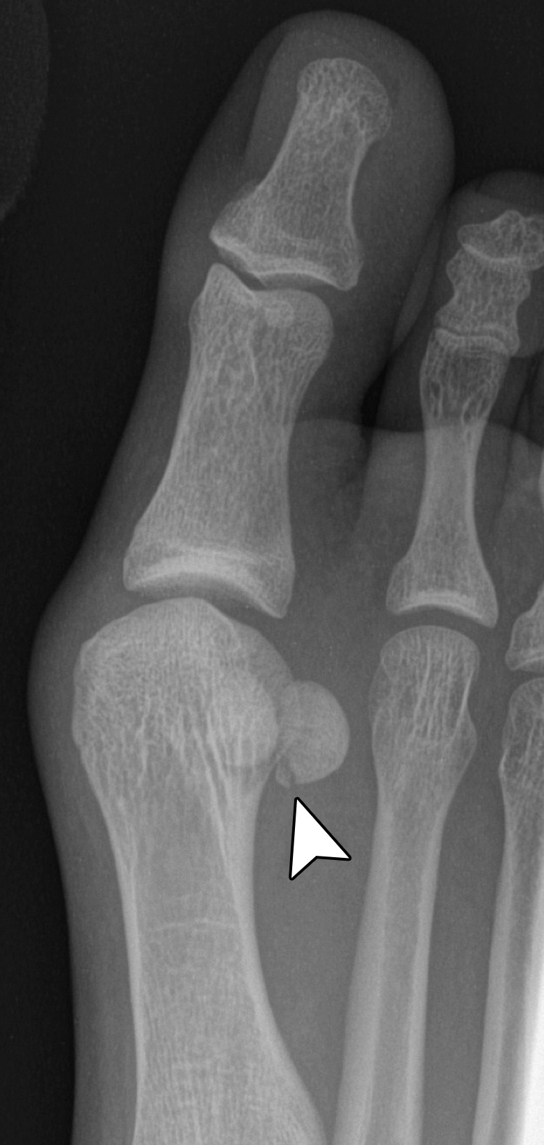
Sesamoid fracture. (a, b) Medial sesamoid fracture in a 32-year-old man after a fall. Axial T2-weighted FS (a) and sagittal PDW (b) images show an irregular cleft through the medial sesamoid with edema at the bony fragments and mild displacement likely caused by a fracture (solid white arrow). An intact medial SPL is apparent (dotted arrow). Differentiating a bipartite sesamoid from a fracture can be challenging. Irregular margins, displacement of the bony components, and a clear history of trauma are more suggestive of a fracture. Radiographs can be useful in assessment and follow-up, but radiographs were not available in this patient. L = lateral sesamoid. (c) Dorsoplantar radiograph in a 49-year-old woman with trauma to the medial sesamoid shows a cleft at the medial sesamoid with slightly irregular margins and no rounded waist (dashed circle). These imaging features are more suggestive of a fracture. (d, e) Dorsoplantar radiograph (d) obtained at 1-year follow-up and axial CT image (e) show interval osseous bridging due to healing. (f) Medial oblique radiograph in a 24-year-old woman shows an irregular fracture of the proximal lateral sesamoid (arrowhead) associated with a dislocation at the first MTPJ due to a motor vehicle accident.

Sesamoid fracture. (a, b) Medial sesamoid fracture in a 32-year-old man after a fall. Axial T2-weighted FS (a) and sagittal PDW (b) images show an irregular cleft through the medial sesamoid with edema at the bony fragments and mild displacement likely caused by a fracture (solid white arrow). An intact medial SPL is apparent (dotted arrow). Differentiating a bipartite sesamoid from a fracture can be challenging. Irregular margins, displacement of the bony components, and a clear history of trauma are more suggestive of a fracture. Radiographs can be useful in assessment and follow-up, but radiographs were not available in this patient. L = lateral sesamoid. (c) Dorsoplantar radiograph in a 49-year-old woman with trauma to the medial sesamoid shows a cleft at the medial sesamoid with slightly irregular margins and no rounded waist (dashed circle). These imaging features are more suggestive of a fracture. (d, e) Dorsoplantar radiograph (d) obtained at 1-year follow-up and axial CT image (e) show interval osseous bridging due to healing. (f) Medial oblique radiograph in a 24-year-old woman shows an irregular fracture of the proximal lateral sesamoid (arrowhead) associated with a dislocation at the first MTPJ due to a motor vehicle accident.