. 2020 May 15;40(4):1107–1124. doi: 10.1148/rg.2020190145

Figure 16a.

Extensor tendon injury in a 70-year-old woman after resection of dorsal first MTPJ osteophytes (cheilectomy). Coronal PDW (a) and sagittal T2-weighted FS (b) images show partial tearing of the extensor tendons at the level of the metatarsal (MT) head, most marked at the EHB (solid white arrow) near the proximal phalanx insertion, with less marked changes at the EHL with an intact distal phalanx (DP) insertion (arrowheads). The dorsal medial sagittal band (black arrow) is partly torn with first MTPJ degeneration and subchondral cysts, along with tearing of the plantar plate complex distally (dotted arrow).

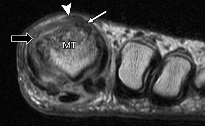
Extensor tendon injury in a 70-year-old woman after resection of dorsal first MTPJ osteophytes (cheilectomy). Coronal PDW (a) and sagittal T2-weighted FS (b) images show partial tearing of the extensor tendons at the level of the metatarsal (MT) head, most marked at the EHB (solid white arrow) near the proximal phalanx insertion, with less marked changes at the EHL with an intact distal phalanx (DP) insertion (arrowheads). The dorsal medial sagittal band (black arrow) is partly torn with first MTPJ degeneration and subchondral cysts, along with tearing of the plantar plate complex distally (dotted arrow).