. 2020 May 15;40(4):1107–1124. doi: 10.1148/rg.2020190145

Figure 17b.

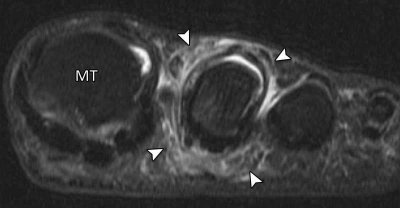
Collateral ligament tears and predislocation syndrome. Axial PDW (a) and coronal T2-weighted FS (b) images show hallux valgus with partial tears of the medial (solid arrow) and lateral (dotted arrow) collateral ligaments with first MTPJ degenerative changes. Capsular thickening and surrounding edema (arrowheads) are seen at the second MTPJ. These imaging features are consistent with adhesive capsulitis, which in association with hallux valgus is termed predislocation syndrome. As the name implies, if the underlying biomechanical abnormality is not corrected, the second MTPJ plantar plate will tear, resulting in instability and eventual dislocation. MT = first metatarsal.

Collateral ligament tears and predislocation syndrome. Axial PDW (a) and coronal T2-weighted FS (b) images show hallux valgus with partial tears of the medial (solid arrow) and lateral (dotted arrow) collateral ligaments with first MTPJ degenerative changes. Capsular thickening and surrounding edema (arrowheads) are seen at the second MTPJ. These imaging features are consistent with adhesive capsulitis, which in association with hallux valgus is termed predislocation syndrome. As the name implies, if the underlying biomechanical abnormality is not corrected, the second MTPJ plantar plate will tear, resulting in instability and eventual dislocation. MT = first metatarsal.