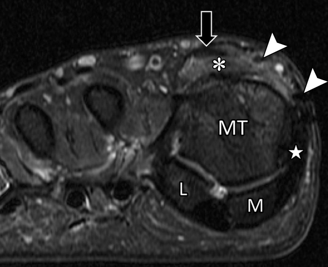
Figure 18b.
Treatment for hallux valgus. Axial PDW (a), coronal T2-weighted FS (b), and sagittal PDW (c) images in a 37-year-old man with reduced extension at the right first MTPJ who had hallux valgus corrective surgery 17 years earlier and hardware removed 1 year before. Postoperative changes and metallic artifacts are seen at the first metatarsal (MT) and proximal phalanx (PP) because of osteotomies. There is thickening of the medial collateral ligament (white arrow) and MTSL (☆) with postsurgical fibrosis appearing as low signal intensity along an attenuated medial sagittal band (arrowheads). Lateralization and tendinosis of the extensor tendons (black arrow) with underlying synovitis, fibrosis, and capsulitis (*) lead to reduced extension. The medial SPL (dotted arrow) shows only mild degeneration. L = lateral sesamoid, M = medial sesamoid.

