. 2020 May 15;40(4):1107–1124. doi: 10.1148/rg.2020190145

Figure 19b.

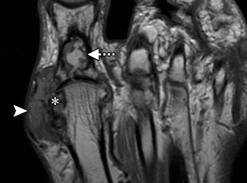
Gouty arthropathy at the first MTPJ in a 73-year-old man with left hallux valgus and first MTPJ pain and swelling. Coronal T2-weighted FS (a) and axial PDW (b) images show first MTPJ degenerative changes with a heterogeneous-signal-intensity soft-tissue mass medially (arrowheads) and bony erosion at the first metatarsal head (*). The underlying medial collateral ligament is poorly visualized, and there is a large proximal phalangeal cyst (dotted arrow) extending from the joint. These findings are compatible with a gouty tophus and intra-articular involvement.

Gouty arthropathy at the first MTPJ in a 73-year-old man with left hallux valgus and first MTPJ pain and swelling. Coronal T2-weighted FS (a) and axial PDW (b) images show first MTPJ degenerative changes with a heterogeneous-signal-intensity soft-tissue mass medially (arrowheads) and bony erosion at the first metatarsal head (*). The underlying medial collateral ligament is poorly visualized, and there is a large proximal phalangeal cyst (dotted arrow) extending from the joint. These findings are compatible with a gouty tophus and intra-articular involvement.