. 2020 May 15;40(4):1107–1124. doi: 10.1148/rg.2020190145

Figure 1b.

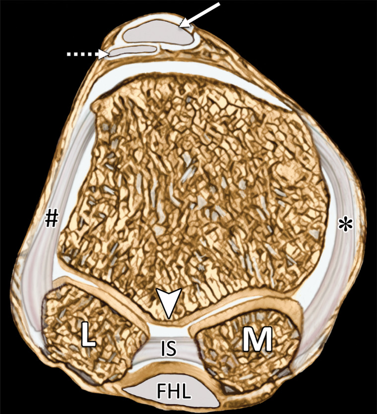
First MTPJ anatomy. Sagittal (a) and coronal cross-sectional (b) drawings (plane in b represented by the dotted line in a) show how the medial SPL (★), lateral SPL (not shown), and lateral (#) and medial (*) MTSLs secure the lateral (L) and medial (M) sesamoids. These ligaments form a plantar plate complex with the joint capsule, ISL, and musculotendinous structures. At the dorsal first MTPJ, the EHB (dotted arrow) attaches to the proximal phalanx (PP) and lies deep and slightly lateral to the EHL (solid arrow). The first metatarsal crest (arrowhead in b) is shown between the grooved sesamoid facets. FHL = flexor hallucis longus, IS = intersesamoid ligament, MT = metatarsal.

First MTPJ anatomy. Sagittal (a) and coronal cross-sectional (b) drawings (plane in b represented by the dotted line in a) show how the medial SPL (★), lateral SPL (not shown), and lateral (#) and medial (*) MTSLs secure the lateral (L) and medial (M) sesamoids. These ligaments form a plantar plate complex with the joint capsule, ISL, and musculotendinous structures. At the dorsal first MTPJ, the EHB (dotted arrow) attaches to the proximal phalanx (PP) and lies deep and slightly lateral to the EHL (solid arrow). The first metatarsal crest (arrowhead in b) is shown between the grooved sesamoid facets. FHL = flexor hallucis longus, IS = intersesamoid ligament, MT = metatarsal.