. 2020 May 15;40(4):1107–1124. doi: 10.1148/rg.2020190145

Figure 20a.

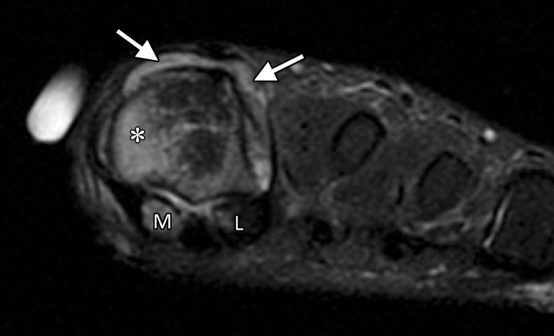
Rheumatoid arthritis at the first MTPJ in a 32-year-old woman with known rheumatoid arthritis and first toe pain and stiffness. Coronal T2-weighted FS (a) and axial T1-weighted (b) images show prominent bone marrow edema at the first MTPJ, mainly involving the metatarsal head (*) as well as the medial (M) and lateral (L) sesamoids. These imaging findings are associated with joint effusion and synovial hypertrophy (solid arrows). An erosion is seen along the lateral aspect of the metatarsal head (dotted arrow). The constellation of findings suggests an inflammatory arthropathy.

Rheumatoid arthritis at the first MTPJ in a 32-year-old woman with known rheumatoid arthritis and first toe pain and stiffness. Coronal T2-weighted FS (a) and axial T1-weighted (b) images show prominent bone marrow edema at the first MTPJ, mainly involving the metatarsal head (*) as well as the medial (M) and lateral (L) sesamoids. These imaging findings are associated with joint effusion and synovial hypertrophy (solid arrows). An erosion is seen along the lateral aspect of the metatarsal head (dotted arrow). The constellation of findings suggests an inflammatory arthropathy.