. 2020 May 15;40(4):1107–1124. doi: 10.1148/rg.2020190145

Figure 21a.

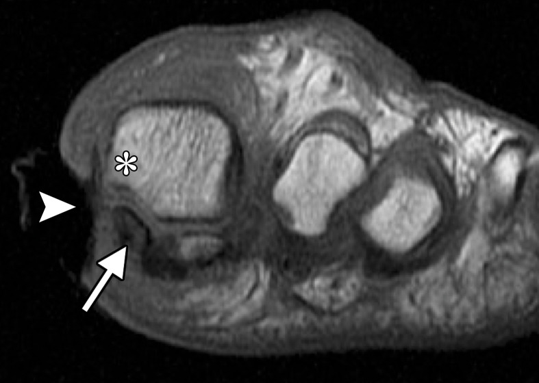
Methicillin-resistant Staphylococcus aureus (MRSA) osteomyelitis at the first MTPJ in a 70-year-old woman with a diabetic wound at the medial first toe. Coronal T1-weighted (a) and T2-weighted (b) FS images show a large soft-tissue ulcer along the medial aspect of the first MTPJ (arrowhead). There is irregularity and loss of the normal T1-weighted marrow signal at the medial sesamoid (arrow) with edema at the sesamoid and medial metatarsal head (*). These changes are suspicious for osteomyelitis. Soft-tissue swelling and edema around the first digit with a small first MTPJ effusion are also noted.

Methicillin-resistant Staphylococcus aureus (MRSA) osteomyelitis at the first MTPJ in a 70-year-old woman with a diabetic wound at the medial first toe. Coronal T1-weighted (a) and T2-weighted (b) FS images show a large soft-tissue ulcer along the medial aspect of the first MTPJ (arrowhead). There is irregularity and loss of the normal T1-weighted marrow signal at the medial sesamoid (arrow) with edema at the sesamoid and medial metatarsal head (*). These changes are suspicious for osteomyelitis. Soft-tissue swelling and edema around the first digit with a small first MTPJ effusion are also noted.