. 2020 May 15;40(4):1107–1124. doi: 10.1148/rg.2020190145

Figure 5b.

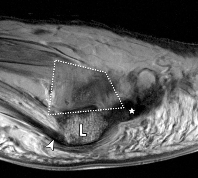
Normal anatomy of the first MTSLs. Coronal (a) and lateral sagittal (b) PDW images (2000/35) depict how the lateral (#) and medial (*) MTSLs secure the lateral (L) and medial (M) sesamoids. The MTSLs are components of the plantar plate complex and attach to the sesamoids in close relation to the FHB tendons. The insertion of the lateral head of the FHB tendon can be seen (arrowhead). The lateral MTSL inserts adjacent to the conjoint adductor hallucis tendon (dotted arrow), which resists varus stress. The medial MTSL inserts adjacent to the abductor hallucis tendon (solid arrow), which resists valgus stress. The rhomboid lateral MTSL (white dotted line in b) blends with the capsule and lateral SPL (☆). Ext = extensor tendons.

Normal anatomy of the first MTSLs. Coronal (a) and lateral sagittal (b) PDW images (2000/35) depict how the lateral (#) and medial (*) MTSLs secure the lateral (L) and medial (M) sesamoids. The MTSLs are components of the plantar plate complex and attach to the sesamoids in close relation to the FHB tendons. The insertion of the lateral head of the FHB tendon can be seen (arrowhead). The lateral MTSL inserts adjacent to the conjoint adductor hallucis tendon (dotted arrow), which resists varus stress. The medial MTSL inserts adjacent to the abductor hallucis tendon (solid arrow), which resists valgus stress. The rhomboid lateral MTSL (white dotted line in b) blends with the capsule and lateral SPL (☆). Ext = extensor tendons.