. 2020 May 15;40(4):1107–1124. doi: 10.1148/rg.2020190145

Figure 8a.

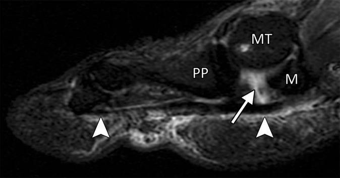
Turf toe in a 28-year-old professional American football player. Arrowheads = FHL, MT = first metatarsal. (a) Sagittal PDW FS image of the right first MTPJ shows complete tearing of the medial SPL (arrow). Additional high-grade tearing of the lateral SPL was apparent (not shown). PP = first proximal phalanx. (b, c) Coronal PDW FS images of the lateral (L) and medial (M) sesamoids (b) and just distal to the sesamoids (c) show the medial SPL tear extending into the ISL (★) and central plantar plate (*). Partial tearing of the medial MTSL (dotted arrow) is also seen. (d) Lateral dynamic dorsiflexed radiograph of both feet shows increased proximal migration of the right medial sesamoid compared to the left medial sesamoid. The medial sesamoids and proximal phalanges are outlined (dashed lines).

Turf toe in a 28-year-old professional American football player. Arrowheads = FHL, MT = first metatarsal. (a) Sagittal PDW FS image of the right first MTPJ shows complete tearing of the medial SPL (arrow). Additional high-grade tearing of the lateral SPL was apparent (not shown). PP = first proximal phalanx. (b, c) Coronal PDW FS images of the lateral (L) and medial (M) sesamoids (b) and just distal to the sesamoids (c) show the medial SPL tear extending into the ISL (★) and central plantar plate (*). Partial tearing of the medial MTSL (dotted arrow) is also seen. (d) Lateral dynamic dorsiflexed radiograph of both feet shows increased proximal migration of the right medial sesamoid compared to the left medial sesamoid. The medial sesamoids and proximal phalanges are outlined (dashed lines).