. 2020 May 15;40(4):1107–1124. doi: 10.1148/rg.2020190145

Figure 9b.

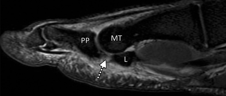
Turf toe in a 24-year-old professional football player. (a, b) Sagittal PDW (a) and sagittal PDW FS (b) images show complete tearing of the lateral SPL (arrow). Complete tearing of the medial SPL was also present (not shown). Slight proximal migration of the lateral (L) and medial (M) sesamoids is noted on the static MR images (a–c). MT = first metatarsal, PP = first proximal phalanx. (c) Axial (long-axis) PDW FS image shows marked edema at the plantar plate complex and lateral (dotted arrow) and medial (solid arrow) SPLs. (d) Lateral dynamic dorsiflexed radiograph of both feet shows proximal medial and lateral sesamoid migration in the right foot because of SPL tearing.

Turf toe in a 24-year-old professional football player. (a, b) Sagittal PDW (a) and sagittal PDW FS (b) images show complete tearing of the lateral SPL (arrow). Complete tearing of the medial SPL was also present (not shown). Slight proximal migration of the lateral (L) and medial (M) sesamoids is noted on the static MR images (a–c). MT = first metatarsal, PP = first proximal phalanx. (c) Axial (long-axis) PDW FS image shows marked edema at the plantar plate complex and lateral (dotted arrow) and medial (solid arrow) SPLs. (d) Lateral dynamic dorsiflexed radiograph of both feet shows proximal medial and lateral sesamoid migration in the right foot because of SPL tearing.