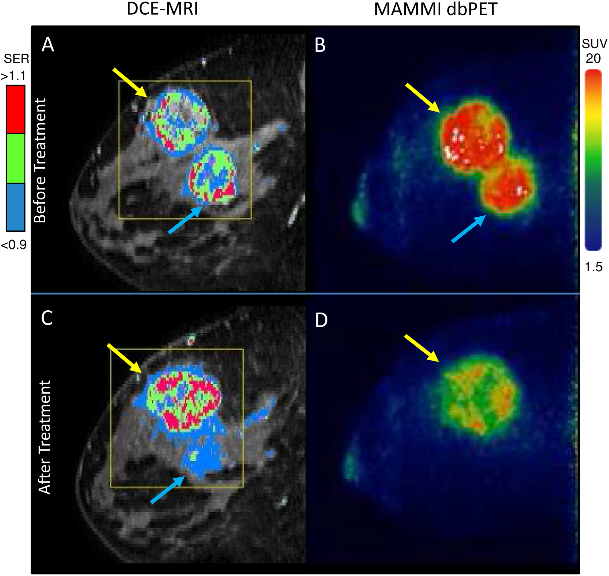
Figure 2.

Breast imaging of a 32 year-old female patient with biopsy confirmed ER+/PR-/HER2- and TN invasive carcinomas in the right breast. A: Before treatment DCE-MRI showing the malignant lesions with the mapping of contrast signal enhancement ratio (SER) and overall FTV at 73.2 cm3. B: Before treatment MAMMI dbPET imaging with FDG confirmed MRI findings, showing high FDG avidity in ER+ (blue arrow, SUVmax = 19.2) and TN (yellow arrow, SUVmax = 19.5) tumors. C: At week 3, DCE-MRI showed residual disease in the ER+ tumor and progression of the TN tumor with the FTV at 89.5 cm3, whereas D: At week 4, MAMMI dbPET showed a complete resolution of FDG uptake in the ER+ tumor and reduction of SUVmax by 22% in the TN tumor.
