Figure 8:

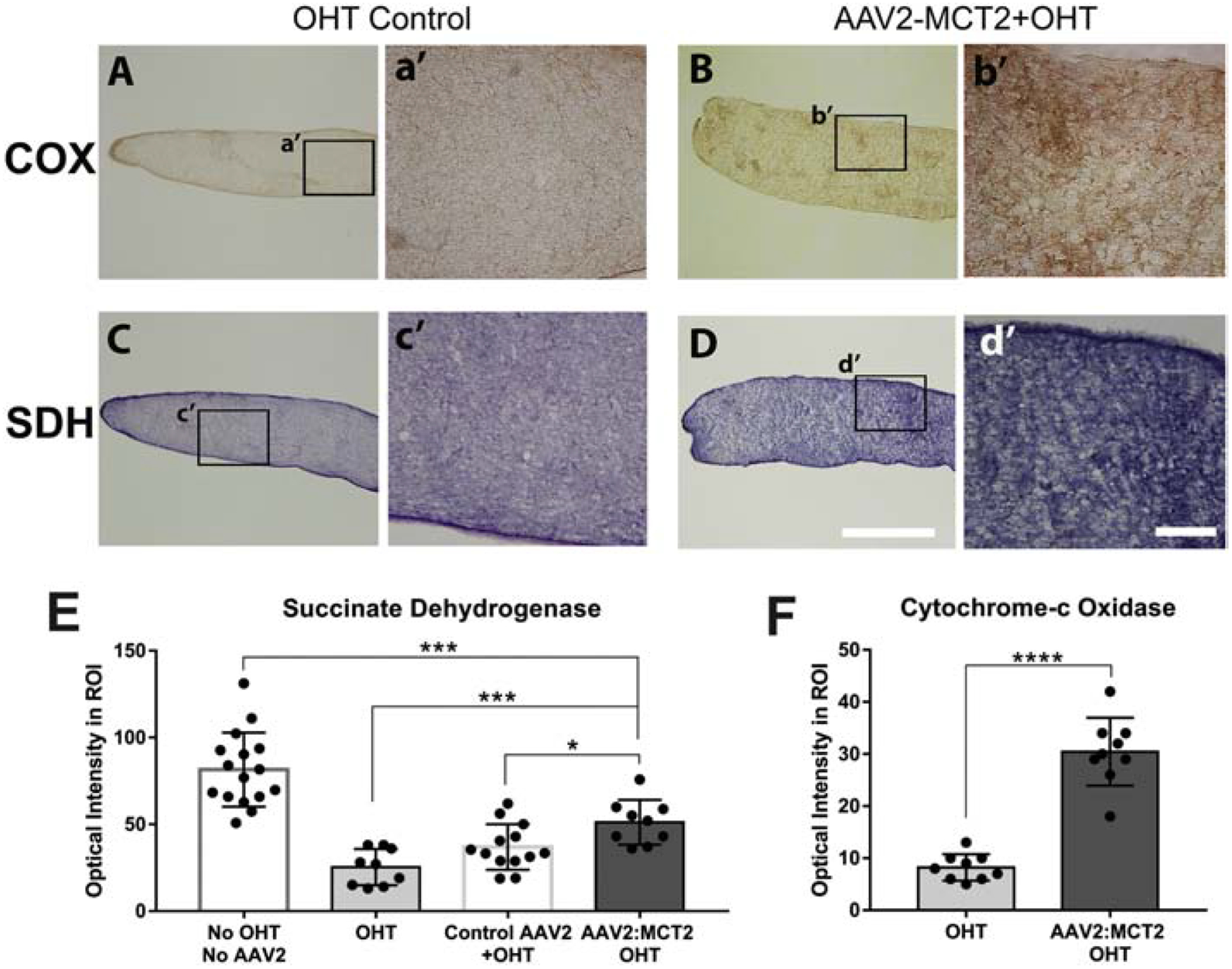
Cytochrome-c oxidase (COX) and succinate dehydrogenase (SDH) activity in OHT optic nerves. A-B. COX histochemistry in OHT mouse ON (A), and OHT with AAV2:MCT2 injection (B). Higher magnification insets of OHT control (a’) and AAV2:MCT2 + OHT (b’) show greater DAB intensity after OHT with AAV2:MCT2. Quantification of histochemistry is shown in F. Scale bar=100μm. C-D. SDH histochemistry in OHT mouse ON (C) and OHT with AAV2:MCT2 injection (D). Scale bar=1000μm. Higher magnification insets of OHT control (c’) and AAV2:MCT2 + OHT (d’) indicate greater nitroblue tetrazolium intensity after OHT with AAV2:MCT2. E. SDH activity is significantly higher in AAV2:MCT2 with OHT ON (n=9 sections across 3 mice) compared to both OHT alone (n=9 sections across 3 mice), and OHT ON injected with control AAV2 (AAV2:GFP) (n=13 sections across 3 mice; p<0.001 and p<0.05, respectively). SDH activity is significantly lower in OHT animals injected with AAV2:MCT2 than in control mice (no OHT or AAV2, n=16 sections across 3 mice). F. COX activity is significantly higher in OHT mouse ON injected with AAV2:MCT2 (n=9 sections across 3 mice) compared to OHT alone (n=9 sections across 3 mice; p<0.0001). Error bars are SD.
