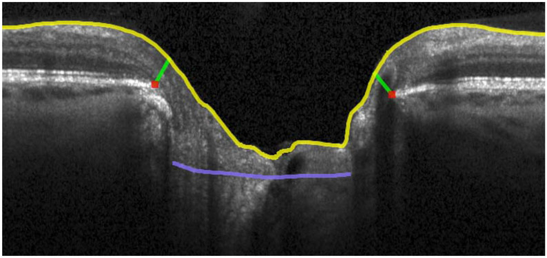
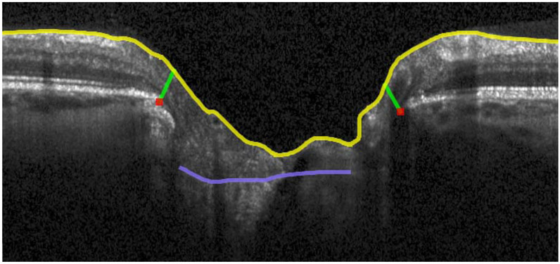
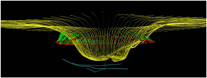
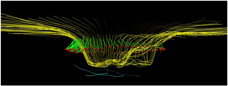
Figure 1.
OCT radial scans of the ONH illustrating changes in MRW, MCD and ALCSD between pre- and post-trabeculectomy scans. 1a: B-scan from the timepoint preceding trabeculectomy. 1b: B-scan from the timepoint following trabeculectomy. 1c: 3D reconstruction of ILM, BMO, MRW and ALCS pre-trabeculectomy. 1d: 3D reconstruction of ILM, BMO, MRW and ALCS post-trabeculectomy. There is a statistically significant improvement in all these ONH parameters which corresponds to a clinical impression of reduced "cupping". Yellow lines: ILM; red dots: BMO; light blue lines: anterior lamina cribrosa surface; green lines: MRW.
MCD: mean cup depth; ALCS: anterior lamina cribrosa surface; ALCSD: anterior lamina cribrosa surface depth; MRW: minimum rim width. BMO: Bruch's membrane opening.


