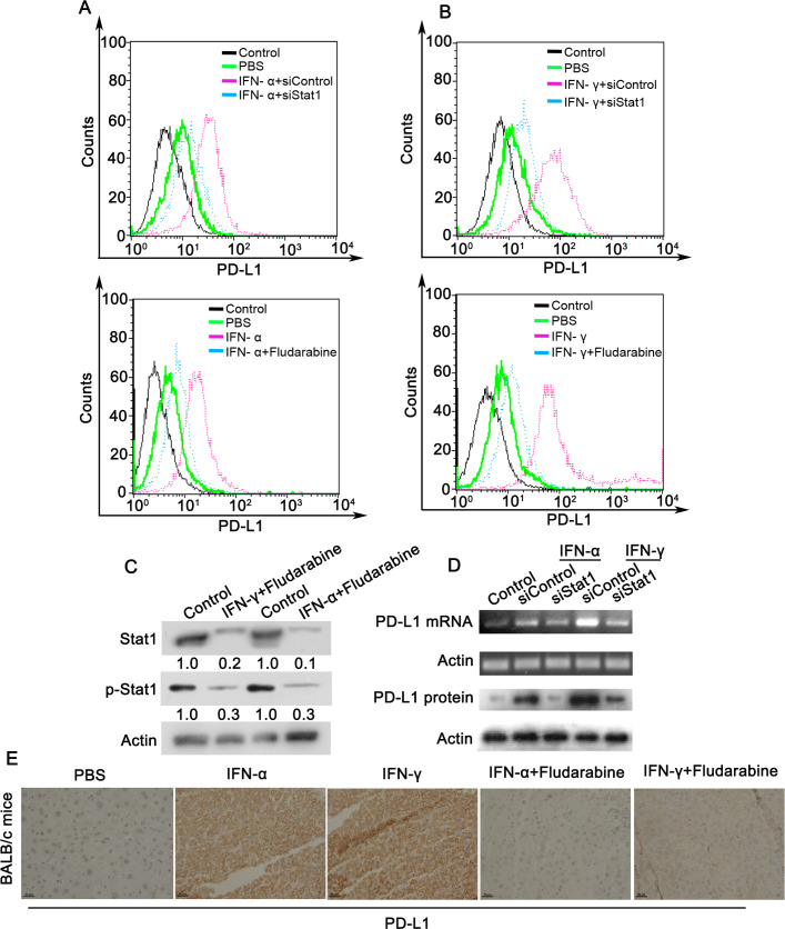
Fig 4. Inhibition of Stat1 abrogates IFN-α/γ-induced upregulation of PD-L1.
(A and B) Cell membrane PD-L1 levels were detected by flow cytometry in L02 cells co-treated with 80 U/ml IFN-α (A) or 50 U/ml IFN-γ (B), and Stat1 siRNA or fludarabine (5 μg/ml) for 48 h. Cells stained with control IgG served as a negative control. (C and D) L02 cells co-treated with 80 U/ml IFN-α or 50 U/ml IFN-γ, with or without fludarabine (5 μg/ml) or Stat1 siRNA for 48 h. Stat1 and phosphorylated Stat1 (p-Stat1) in cells co-treated with or without (control) fludarabine were determined by western blot (C). The mRNA and protein levels of PD-L1 were analyzed using real-time PCR and western blotting, respectively (D). (E) BALB/c mice were treated with PBS, IFN-α, and IFN–γ, and fludarabine as described in Materials and Methods. IHC analysis was performed for detection of PD-L1 expression levels in mouse livers. Scale bars, 50 μm. The experiments were performed twice with similar results.

