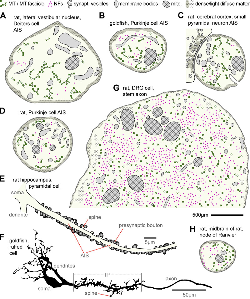
Figure 2.
Ultrastructural features at the AIS, stem axon, and NoRs. (A) AIS of a giant cell of Deiters in the rat lateral vestibular nucleus (Palay et al., 1968). (B) AIS of a goldfish Purkinje cell (Matsumura and Kohno, 1991). (C) AIS of a small pyramidal neuron of the rat cerebral cortex (Peters et al., 1968) bearing an input synapse (IS), which is symmetric (thin postsynaptic undercoat) with flat pleiotropic vesicles. (D) AIS of a rat Purkinje cell (Chan-Palay, 1972). (E) Reconstruction from a TEM series of a pyramidal cell AIS with numerous presynaptic boutons and small spines in the rat hippocampus (redrawn from Kosaka, 1980a). (F) Initial unmyelinated portion (IP; proximal part of which is the AIS; see Table S1) of a ruffed cell in the frog olfactory bulb with numerous pre- and postsynaptic spines (redrawn from Kosaka and Hama, 1979b). (G) Stem axon of a DRG cell in rat (Nakazawa and Ishikawa, 1995). (H) NoR in the midbrain of rat (Peters, 1966). Note that NFs are sparse in AISs and that MTs are not fasciculated in NoRs. Best guesses were made with respect to structures drawn; scales in most original photos were given as magnifications (unfortunately meaningless in PDF format) and were estimated here by measuring MT diameters; the suggested black scale bar for A–D, G, and H represents 500 nm. Publications providing further examples of stem axons or NoRs are provided in the main text; for further AIS descriptions, see Table S1.

