Figure 4.
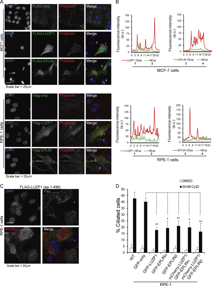
LUZP1 and EPLIN have actin stabilization roles. (A) IF analysis of MCF-7 (top panel) and RPE-1 (bottom panel) cells transiently transfected with a control empty FLAG vector or plasmids to express FLAG-LUZP1 or FLAG-EPLINβ. The fusion proteins were detected with a FLAG antibody. The actin cytoskeleton was stained with fluorophore-conjugated phalloidin, and DNA was stained with DAPI. (B) The graphs show fluorescence intensity of phalloidin in MCF-7 and RPE-1 cells overexpressing LUZP1 or EPLIN and neighboring cells measured along the lines indicated in A. NExp, nonexpressing; OExp, overexpressing. (C) IF analysis of RPE-1 cells transiently transfected with a FLAG-LUZP1 (aa 1–496) construct. The actin cytoskeleton was stained with fluorophore-conjugated phalloidin, and DNA was stained with DAPI. (D) Bar graph shows the mean percentage of ciliated cells (n > 200 cells per sample, three independent experiments) in the indicated cell lines treated with DMSO or 50 nM cytochalasin D (CytD). Error bars indicate SD. *, P < 0.05; **, P < 0.01 (Student’s two-tailed t test).

