Figure S4.
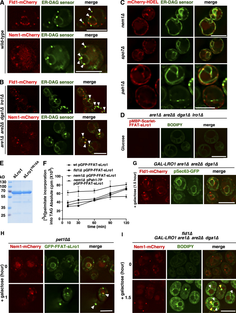
Data associated with Figs. 4, 5, 6, 7, and 8. Enrichment of DAG at Fld1 and Nem1 ER subdomains. (A) ER subdomains marked by Fld1 and Nem1 show enrichment of DAG. WT cells expressing Fld1-mCherry or Nem1-mCherry and coexpressing the GFP-tagged ER-DAG sensor were grown in SC media to early stationary phase and imaged live. White arrowheads indicate colocalization of the ER-DAG sensor with ER punctae marked with Fld1 or Nem1. Scale bars, 5 µm. (B) Fld1- and Nem1-marked ER subdomains show enrichment of DAG in LD-deficient cells. 4ΔKO cells expressing Fld1-mCherry or Nem1-mCherry and coexpressing the ER-DAG sensor were grown as described in A. White arrowheads indicate colocalization of the ER-DAG sensor with punctae marked by Fld1 or Nem1. Scale bar, 5 µm. (C) Lack of Nem1, Spo7, or Pah1 results in uniform distribution of the ER-DAG sensor. nem1Δ, spo7Δ, or pah1Δ mutant cells expressing the ER-DAG sensor were grown as described in A. ER was visualized by mCherry-HDEL. Scale bar, 5 µm. (D) BODIPY staining of 4ΔKO cells expressing MBP-Scarlet-FFAT-sLro1. Cells were grown in glucose media to mid-log phase. Scale bar, 5 µm. (E) Coomassie blue staining of an SDS-PAGE gel showing purified WT and mutant sLro1. (F) Rate of TAG synthesis. Time-dependent incorporation of [3H]palmitic acid into TAG in the indicated yeast mutant cells. Cells were labeled and collected at 10-, 30-, 60-, 90-, and 120-min time points. Data are means ± SD of three independent experiments. (G) An ER membrane protein that is not involved in LD biogenesis does not show enrichment at Fld1-marked ER subdomains. The ER protein Sec63 does not become enriched at Fld1 sites during induction of LD biogenesis. GAL-LRO1 3ΔKO cells expressing Fld1-mCherry and coexpressing Sec63-GFP were grown in raffinose media and switched to galactose-containing media for 1.5 h. Scale bar, 5 µm. (H) Lack of Pet10 does not affect Nem1 localization and recruitment of TAG-synthase. The recruitment of GFP-FFAT-sLro1 to Nem1 sites is not affected in pet10Δ mutant cells. pet10Δ mutant cells expressing Nem1-mCherry and coexpressing GFP-FFAT-sLro1 from a galactose-inducible promoter were shifted to galactose media for the indicated period of time. Arrowheads denote colocalization of sLro1 with Nem1 foci. Scale bar, 5 µm. (I) Neutral lipids accumulate in the ER membrane in seipin mutant cells. fld1Δ mutant cells (fld1Δ GAL-LRO1 3ΔKO) expressing Nem1-mCherry were grown in raffinose media and transferred to galactose-containing media for the indicated period of time, stained with BODIPY, and imaged. White arrowheads indicate BODIPY accumulation in the ER. Yellow arrows indicate BODIPY punctae that do not colocalize with Nem1 foci. Scale bar, 5 µm.

