
Figure 4.
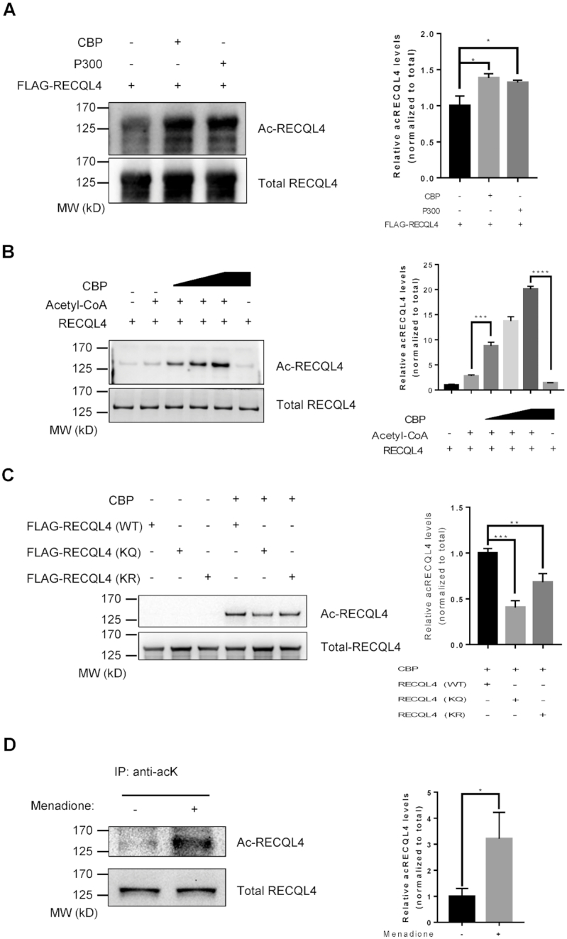
RECQL4 is an acetylated protein and its acetylation is stimulated by oxidative stress. (A) RECQL4 acetylation in transiently transfected U2OS cells with 3xFLAG-tagged RECQL4, p300 or CBP, treated with 2 μM TSA (trichostatin A) and 5 mM NAM (nicotinamide). Immunoprecipitated RECQL4 proteins were then detected by Western blot with anti-acetylated lysine antibody. A representative gel is shown on the left. The graph on the right shows quantification of relative acetylated RECQL4 levels normalized to total RECQL4. Data are presented as mean ± SD from two independent experiments. *P < 0.05, using unpaired two-tailed Student's t test. (B) Acetylation of RECQL4 by CBP in an in vitro acetylation assay. Recombinant RECQL4 (1 μg), Acetyl-CoA (2 mM), and different amounts of recombinant CBP (0.1, 0.2, 0.5 μg) were incubated at 30°C for 1 h. Acetylated RECQL4 proteins were detected with anti-acetylated lysine antibody (top). Total amounts of RECQL4 were assessed with Coomassie blue staining (bottom). The graph on the right shows quantification of relative acetylated RECQL4 levels normalized to total RECQL4. Data are shown as mean ± SD from three independent experiments. ***P < 0.001, ****P < 0.0001, using unpaired two-tailed Student's t test. (C) Acetylation of wild type RECQL4 (WT), RECQL4 (KQ) and RECQL4 (KR) mutants by CBP in an in vitro acetylation assay. 3 × FLAG-tagged RECQL4 proteins were purified from normal U2OS cell. FLAG-tagged RECQL4 proteins (1 μg), Acetyl-CoA (2 mM), and recombinant CBP (0.2 μg) were incubated at 30°C for 1 h. Acetylated FLAG-tagged RECQL4 proteins were detected with anti-acetylated lysine antibody. A representative gel is shown on the left. The graph on the right shows quantification of relative acetylated FLAG-tagged RECQL4 protein levels normalized to total. Data are shown as mean ± SD from two independent experiments. **P < 0.01, ***P < 0.001, using unpaired two-tailed Student's t test. (D) Western blot showing increased endogenous acetylated RECQL4 levels in cells treated with 50 μM menadione for 1 h. Endogenous acetylated proteins were immunoprecipitated with anti-acetyl-lysine antibody-conjugated beads. The presence of RECQL4 in the immunoprecipitated complex and acetylation levels in the extract were measured by Western blot with anti-RECQL4 antibody. Input for immunoprecipitation was used as a loading control for total RECQL4 (bottom). A representative gel is shown on the left. The graph on the right shows quantification of relative acetylated RECQL4 levels normalized to total RECQL4. Data are shown as mean ± SD from two independent experiments. *P < 0.05, using unpaired two-tailed Student's t test.