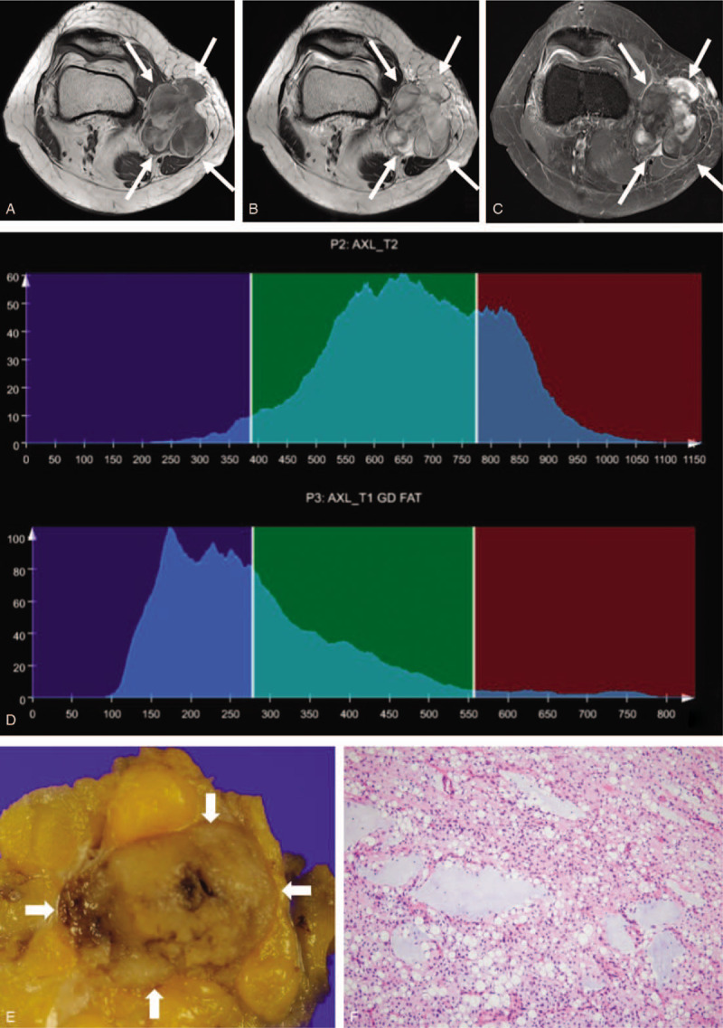
Figure 2.

A 50-year-old woman with myxoid liposarcoma, grade 1. A mass with a lobulated margin in the medial side of knee is hypointense on axial T1-weighted imaging (a), heterogeneously hyperintense on axial T2-weighted imaging (b) and exhibits heterogeneous enhancement on axial fat-suppressed contrast-enhanced T1-weighted imaging (c). Whole-tumor T2 (upper) and contrast-enhanced (CE) T1 (lower) histograms (d) reveal negative T2 skewness (–0.104) and high CE T1 skewness (1.345). Texture features were high T1 standard deviation (108) and low CE T1 difference variance (0.176). These findings suggest low-grade sarcoma. (e) Grossly, the tumor is well circumscribed and shows gelatinous, tan-yellow cut surface. (f) In this microscopic image (hematoxylin-eosin stain; original magnification, x100), the tumor of low cellularity is composed of small spindled or ovoid non-lipogenic tumor cells with scant cytoplasm and lipoblasts in background of abundant myxoid stroma and arborizing capillary vasculature.
