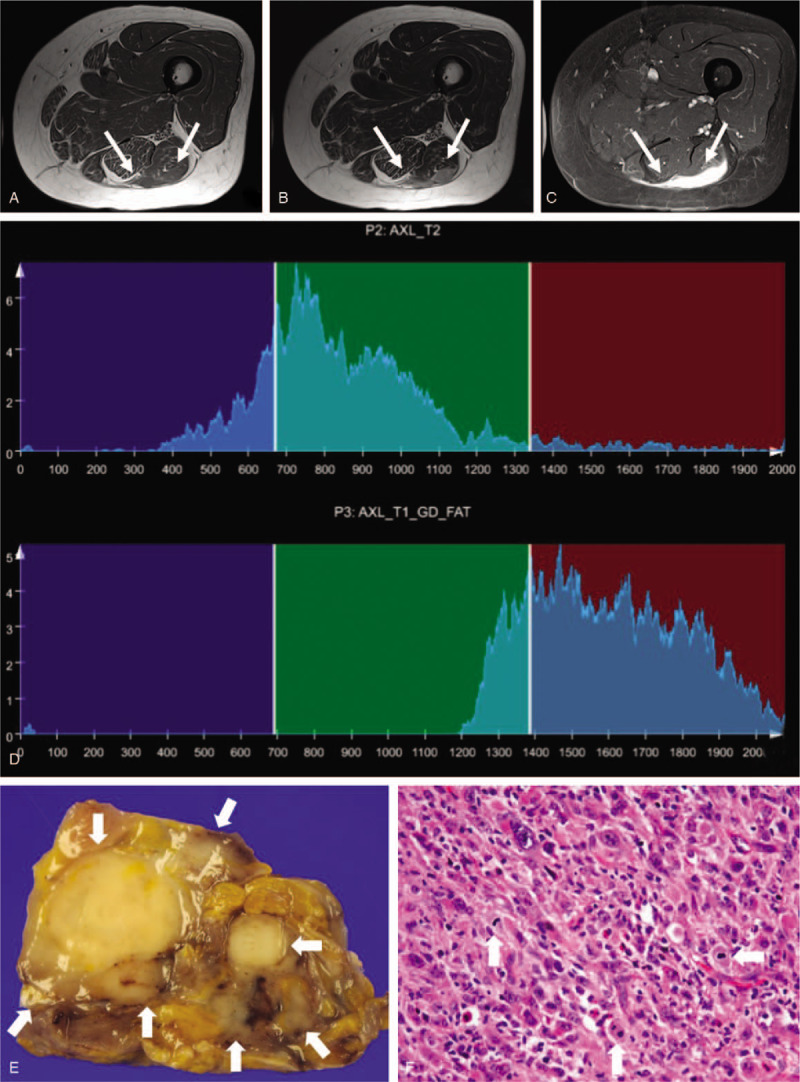
Figure 4.

A 61-year-old woman with undifferentiated sarcoma, grade 3. Axial T1-weighted imaging (a) demonstrates a homogeneously hypointense mass within the posterior thigh. Axial T2-weighted imaging (b) shows relatively homogeneous hypointensity to intermediate signal. The mass shows homogeneous, intense contrast enhancement on axial fat-suppressed contrast-enhanced T1-weighted imaging (c). Whole-tumor T2 (upper) and contrast-enhanced (CE) T1 (lower) histograms (d) reveal high T2 skewness (1.233) and negative CE T1 skewness (-0.529). Texture features are high T2 mean (855.3), high T1 standard deviation (234), and high CE T1 difference variance (0.305). Texture features, except for T2 mean and T1 standard deviation, are compatible with high-grade sarcoma. (e) Grossly, an ill-defined tumor shows a lobulated, firm, tan-white cut surface. (f) In this microscopic image (hematoxylin-eosin stain; original magnification, x400), pleomorphic tumor cells are admixed with abundant chronic inflammatory cells and show frequent mitosis (arrows).
