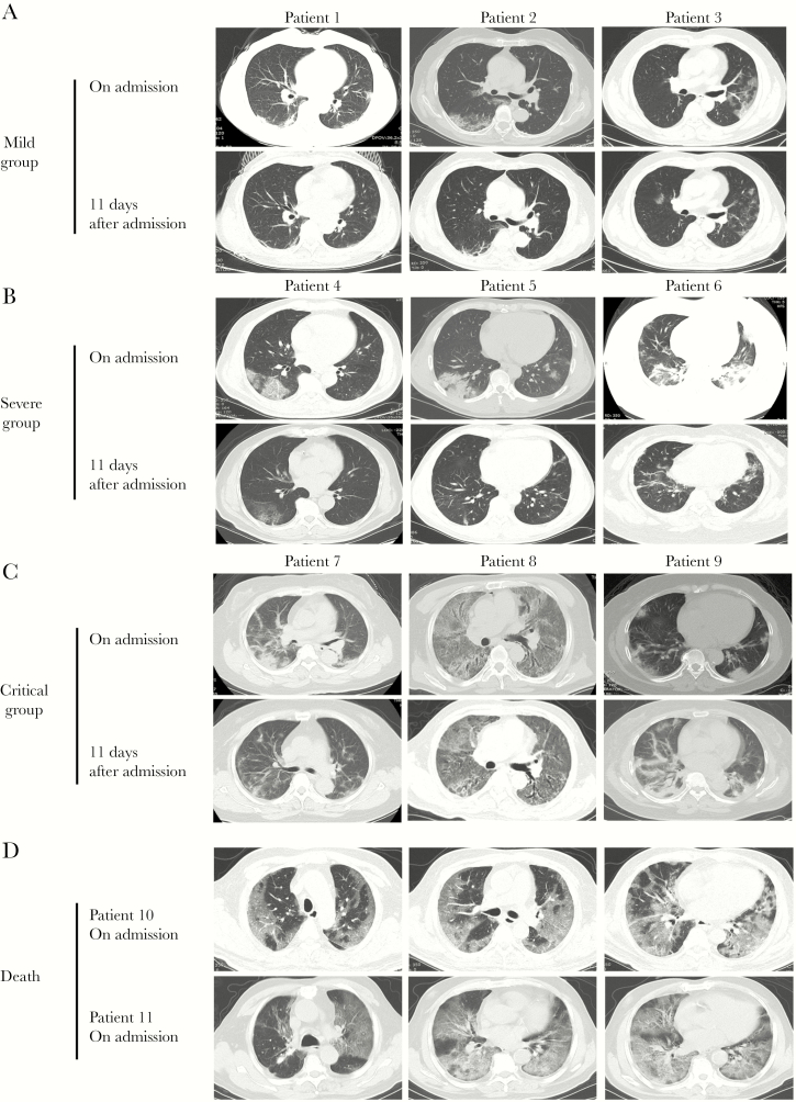
Figure 3.
Chest computed tomography (CT) images of patients with coronavirus 2019 (COVID-19). (A) Transverse chest CT images showed bilateral ground-glass opacity from 3 mild patients 1 week after symptom onset and 11 days after admission. (B) Chest CT images showed subsegmental areas of consolidation from another 3 severe patients 1 week after symptom onset and 11 days after admission. (C) Extensive bilateral pulmonary changes of consolidation were found in the lungs of critical patients. (D) Patients who died in the critical group had more severe lesions in bilateral lungs than those in the mild and severe groups.

