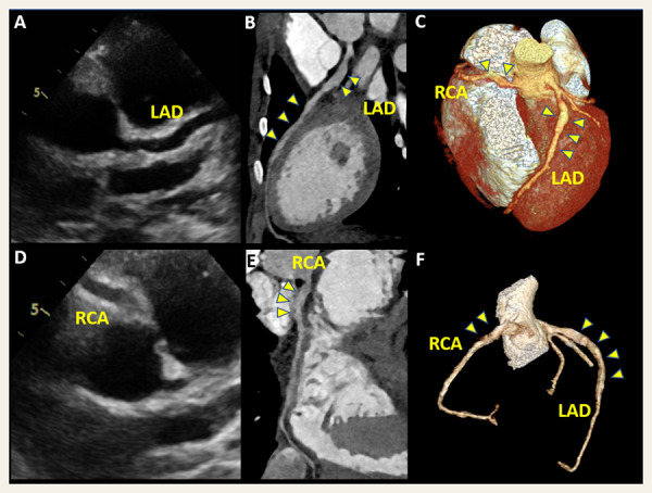
We report the case of a child with coronary artery dilatation in the context of multisystem inflammatory syndrome temporally associated with SARS-CoV-2 infection, without diagnostic criteria for Kawasaki disease.
Although relatively spared by the COVID-19 pandemic, children can present with multisystem inflammatory syndrome temporally associated with SARS-CoV-2 infection (MIS-C), and cardiovascular involvement that shares some similarities with Kawasaki disease (KD). This report describes a case of MIS-C with coronary artery dilatation.
A 10-year-old overweight male was admitted to the intensive care unit (ICU) in mid April 2020 in hypotensive shock with MIS-C after 7 days of fever and gastrointestinal symptoms. No clinical features of KD were found. Recent SARS-CoV-2 infection was proven with positive IgA and IgG serologies, whereas multisite PCRs were negative. Inflammatory parameters were elevated on admission, as were cardiac enzymes [brain natriuretic peptide (BNP) 17 814 ng/L and troponin T 299 ng/L].

Cardiac involvement consisted initially of a mild reduction of left ventricular systolic function [ejection fraction (EF) 40%], with no signs of myocarditis on cardiac magnetic resonance imaging (MRI). By day 8, echocardiography showed left anterior descending (LAD) and right coronary artery (RCA) long segmental dilatations (Panels A and D). Cardiac function had normalized. ECG was unremarkable. The patient was treated with 2 g/kg intravenous immunoglobulin (IVIG), corticosteroids, and anakinra, with favourable clinical evolution and resolution of the inflammatory syndrome.
One month after diagnosis, coronary artery dilatation was confirmed on computed tomography (CT), with long fusiform dilatations: LAD maximal diameter was 6.2 mm (z-score +7.9) and RCA was 4.2 mm (z-score +2.9) (Panels B and E, curved multiplanar reconstruction; Panels C and F, volume rendering). Coronary artery dilatation after SARS-CoV-2 infection is of concern. It probably represents a post-infectious vasculitis and requires further aetiological and follow-up research.
Author contributions: all authors participated in writing and approved the final manuscript. Written consent for publication was obtained from the patient.


