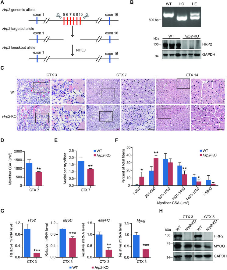
Figure 9.
Muscle regeneration is impaired in Hrp2 KO mice treated with CTX. (A) Schematic outlining the generation of Hrp2 knockout mice using the CRISPR/Cas9 system. The targeting sites of Hrp2 are shown. (B) Hrp2−/− KO male mice were established by breeding Hrp2+/− males and females. The targeted fragment of Hrp2 was amplified by PCR using genomic DNA templates (upper). HRP2 abundance in WT and Hrp2 knockout mice was examined by western blot (lower). HO: homozygous, HE: heterozygous. (C) Representative H&E-stained cross-sections of TA muscles from male WT and Hrp2−/− mice. Sections were obtained from CTX-injured muscles at Days 3, 7 and 14 post-treatment. Boxed areas are enlarged in the panels to their right. Scale bar, 50 μm. (D) Mean myofiber cross-sectional area (CSA) of regenerating muscles in male WT and Hrp2−/− mice (number of fibers counted > 1000) at Day 7 post-CTX-induced injury (n = 4 per group). (E) Numbers of nuclei per myofiber in TA muscle from WT or Hrp2−/− mice at Day 7 post-CTX-induced injury (n = 4 per group). (F) Myofiber CSA distribution in WT and Hrp2−/− mice 7 days after CTX injection. (G) mRNA levels of embryonic MyHC, Myog, MyoD and Hrp2 were analyzed in TA muscle of WT or Hrp2−/− mice at Day 3 post-CTX-induced injury (n = 3 per group). (H) Western blot analysis of MYOG and GAPDH protein levels in TA muscle of WT or Hrp2−/− mice at Days 3 and 5 post-CTX-induced injury. Data are represented as means ± SD of 4 (D–F) or 3 (G) replicates. ** P < 0.01 and *** P < 0.001.

