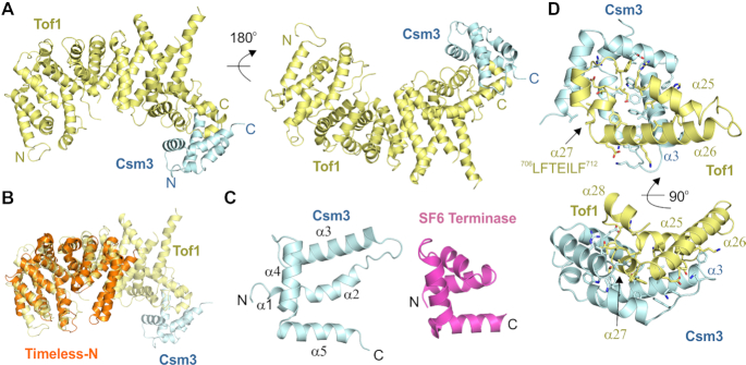
Figure 1.
Crystal structure of the Tof1–Csm3 complex. (A) The complex is shown from two views with Tof1 colored in yellow and Csm3 in cyan. (B) Superposition of an N-terminal fragment of Timeless (PDB 5MQI) onto Tof1. (C) The structure of Csm3 and comparison to a protein with a similar fold, the DNA binding domain of SF6 small terminase (PDB 4ZC3). (D) Details of the interaction between Tof1 and Csm3. Only α25-27 of Tof1 are shown, and interfacial residues are shown in stick representation.

