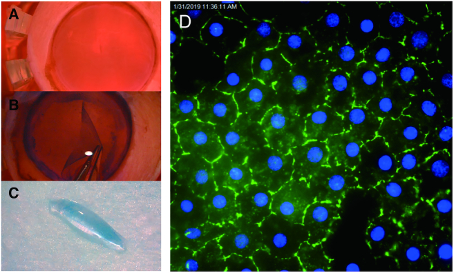
Figure 1.
Preparation of homogeneous tissue monolayers for analysis. (A) Human donor cornea in corneal viewing chamber with Optisol corneal storage media (Bausch & Lomb). (B) Corneal endothelium/Descemet's membrane monolayer being dissected from underlying stromal tissue. (C) Monolayer of corneal endothelial cells assumes a ‘scroll’ shape. This single cell monolayer will be used for sequencing and other analyses. (D) Immunostaining of monolayer of cells with corneal endothelial-specific marker, zonula occudens-1 (ZO-1) (Blue-DAPI).

