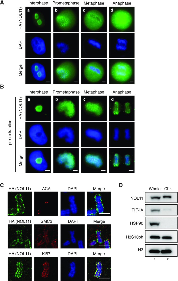
Figute 1.

NOL11 localises at the chromosome periphery. (A–C) Localisation of NOL11 at the PR. HeLa cells that stably expressed FLAG- and HA-tagged NOL11 (FLAG/HA-NOL11 HeLa cells) were fixed with 4% PFA after pre-extraction (B) or without pre-extraction (A) and stained with anti-HA antibodies (NOL11; green) and DAPI (blue). Representative cells in interphase (a), prometaphase (b), metaphase (c), and anaphase (d) are shown. Scale bar, 5 μm. (C) Chromosome spreads prepared from FLAG/HA-NOL11 HeLa cells were co-stained with anti-HA antibodies (NOL11; green) and ACA (red), anti-SMC2 antibodies (red), or anti-Ki67 antibodies (red) and then DAPI (blue). Scale bar, 2 μm. (D) Localisation of NOL11 in the chromosomal fraction of mitotic cells. HeLa cells were synchronised in mitosis and fractionated. Whole-cell extracts (Whole) and chromosomal fractions (Chr.) were immunoblotted with the indicated antibodies. HSP90 is a marker for cytosolic proteins and H3 is a marker for chromosomal proteins. Note that mobility of NOL11 in the chromosomal fraction appears slower than that in the Whole-cell extracts.
