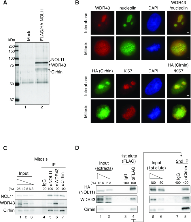
Figure 2.
NOL11, WDR43, and Cirhin comprise the NWC complex. (A) Identification of NOL11-interacting proteins. Extracts prepared from nocodazole-treated HeLa cells (Control) or those stably expressing FLAG/HA-NOL11 were incubated with anti-FLAG antibody-conjugated agarose beads, and bound proteins were eluted using the FLAG peptide. The eluates were subsequently immunoprecipitated with anti-HA antibody-conjugated protein G Sepharose beads. Bound proteins were resolved through SDS-PAGE, visualised by silver staining, and analysed via MS. (B) Localization of WDR43 and Cirhin. Interphase (top row) or mitotic (second row) HeLa cells were co-stained with anti-WDR43 antibodies (red) and anti-Nucleolin antibodies (green), followed by DAPI staining (blue). Alternatively, interphase (third row) or mitotic (bottom row) HeLa cells stably expressing FLAG/HA-Cirhin were co-stained with anti-HA antibodies (green) and anti-Ki67 antibodies (red), followed by DAPI staining (blue). Scale bar, 2 μm. (C) Endogenous interactions among NOL11, WDR43, and Cirhin. Endogenous NOL11, WDR43, or Cirhin was immunoprecipitated from mitotic HeLa cell extracts and then immunoblotted with the indicated antibodies. (D) Complex formation by NOL11, WDR43 and Cirhin. Mitotic extracts prepared from HeLa cells stably expressing FLAG/HA-NOL11 were mixed with normal mouse IgG or anti-FLAG antibody-conjugated agarose beads, and bound proteins were eluted using the FLAG peptides (1st elute). Then, the eluates from anti-FLAG antibody-conjugated agarose beads were immunoprecipitated using anti-Cirhin antibodies (second IP). Proteins were analysed using the indicated antibodies.

