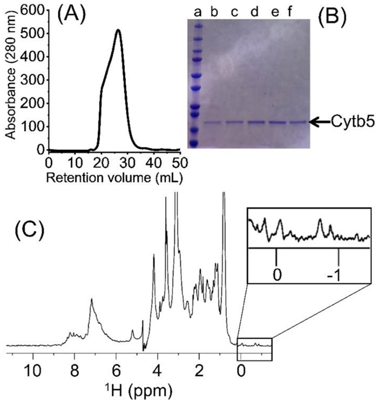
Figure 2.
(A) Size-exclusion chromatogram (SEC) of the extracted cytochrome-b5. (B) SDS-PAGE analysis of the directly extracted cytochrome-b5. Lane a includes the protein markers and lanes b-f the SEC protein fractions. (C) 1H NMR spectrum of the directly extracted cytochrome-b5. Methyl resonances in the upfield region (boxed) indicating the folded conformation of the protein. The protein sample was prepared in 20 mM potassium phosphate buffer pH 7.4, 50 mM NaCl. The NMR spectrum data were collected at 25 °C using a Bruker 500 MHz NMR spectrometer equipped with a TXI probe.

