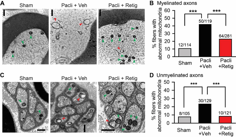
Figure 4.
The effect of early, repeated application of retigabine on the morphological alteration of mitochondria in tibial nerves after paclitaxel. Representative images of swollen mitochondria (red arrows) with vacuoles and small oval mitochondria (green arrows) with intact double membranes in myelinated axons (A) and unmyelinated axons (C) of tibial nerve sections from sham (left panel), paclitaxel plus vehicle (middle panel), and paclitaxel plus retigabine (right panel) groups. Scale bars, 0.5 μm. (B) and (D) Quantification of abnormal mitochondria in both myelinated axons and unmyelinated axons. P = 0.0003, X2 = 16.442 for unmyelinated fibers; P < 0.0001, X2 = 32.213 for myelinated fibers; Chi-square tests. N is the number of fibers tested. Four animals for each group were used. Data were collected 28 days after initial paclitaxel treatment. *, p<0.05; **, P<0.01, ***, P<0.001.

