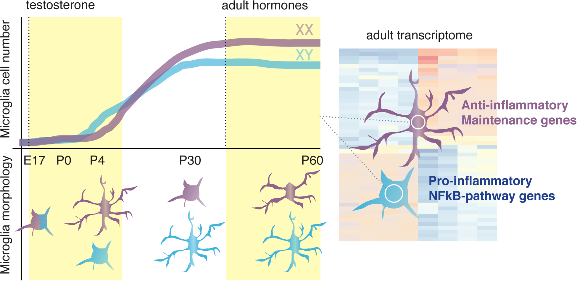
Figure 2. Overview of sex differences in microglia.

An overview of differences in male and female microglia in rodents, highlighting differences in cell number (upper half of graph) and morphology (bottom half of graph) during development and in the adult brain [43], as well as transcriptional differences in the adult rodent [49,50]. Purple and blue microglia represent those from female and male animals, respectively.
