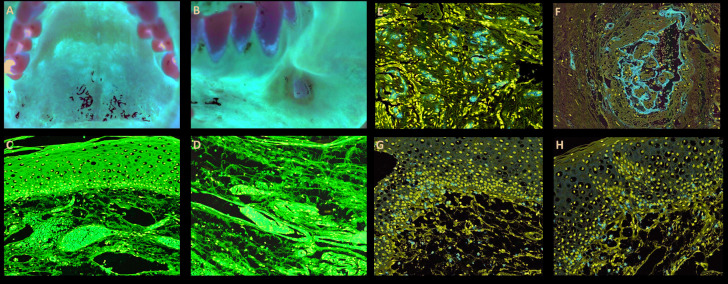
Figure 1.
Clinical and microscopic features of reddish oral lesions in a patient with Covid-19. A: Diffuse reddish lesion in the hard palate. B: Ulcer with ischemic aspect in the buccal mucosa. C: Epithelium demonstrating vacuolization and haemorrhage in the superficial portion of the lamina propria, with hyperaemic vessels. D: Lymphocytic infiltration in the connective tissue and different-sized thrombi. E: CD34 positive expression in thrombi of small vessels. F: Larger thrombi with variable amount of fibrin and endothelial cells positive for CD34. G: CD3 positivity in most inflammatory cells in the connective tissue and basal layer of the epithelium. H: CD8 highlighting lymphocytes in the lamina propria and basal layer of the epithelium.

