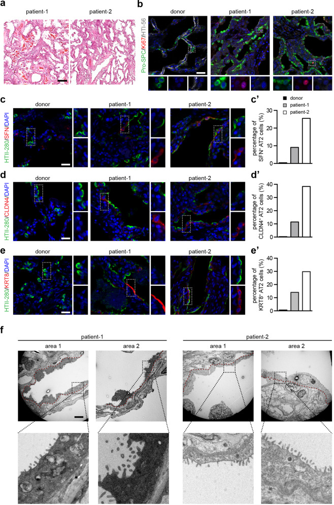
Fig. 1. The proliferation and differentiation of the resident alveolar stem cells in the lungs of two COVID-19 patients.
a H&E staining of the lungs of two COVID-19 patients. b Immunostaining using antibodies against proSPC, Ki67, and HTI-56 of a healthy donor lung and lungs of two COVID-19 patients. c–e′ Immunostaining using antibodies against HTII-280 and SFN (c), HTII-280 and CLDN4 (d), and HTII-280 and KRT8 (e) of a healthy donor lung and the COVID-19 lungs. The percentage of intermediate AT2 cells in total AT2 cells were quantified (c′, d′, and e′). f TEM images of the COVID-19 lungs. The red dashed line indicates the location of the basement membrane. Scale bars, 100 µm (a), 20 µm (b–e), 5 µm (f).

