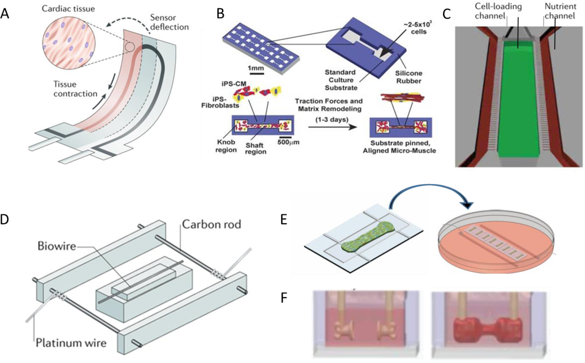
Figure 2. Representative template-guided heart-on-a-chip devices.

Cardiomyocytes can compact A) adhering as muscle thin films on top of 3D printed sensors[57, 62], B) within microgrooves[59], C) along patterned perfusable channels [62, 63], D) around single suture template [62, 64], E) around double elastic force sensors[28], and F) upside down force-sensing posts[32]. Figures are adapted with permission from their original references.
