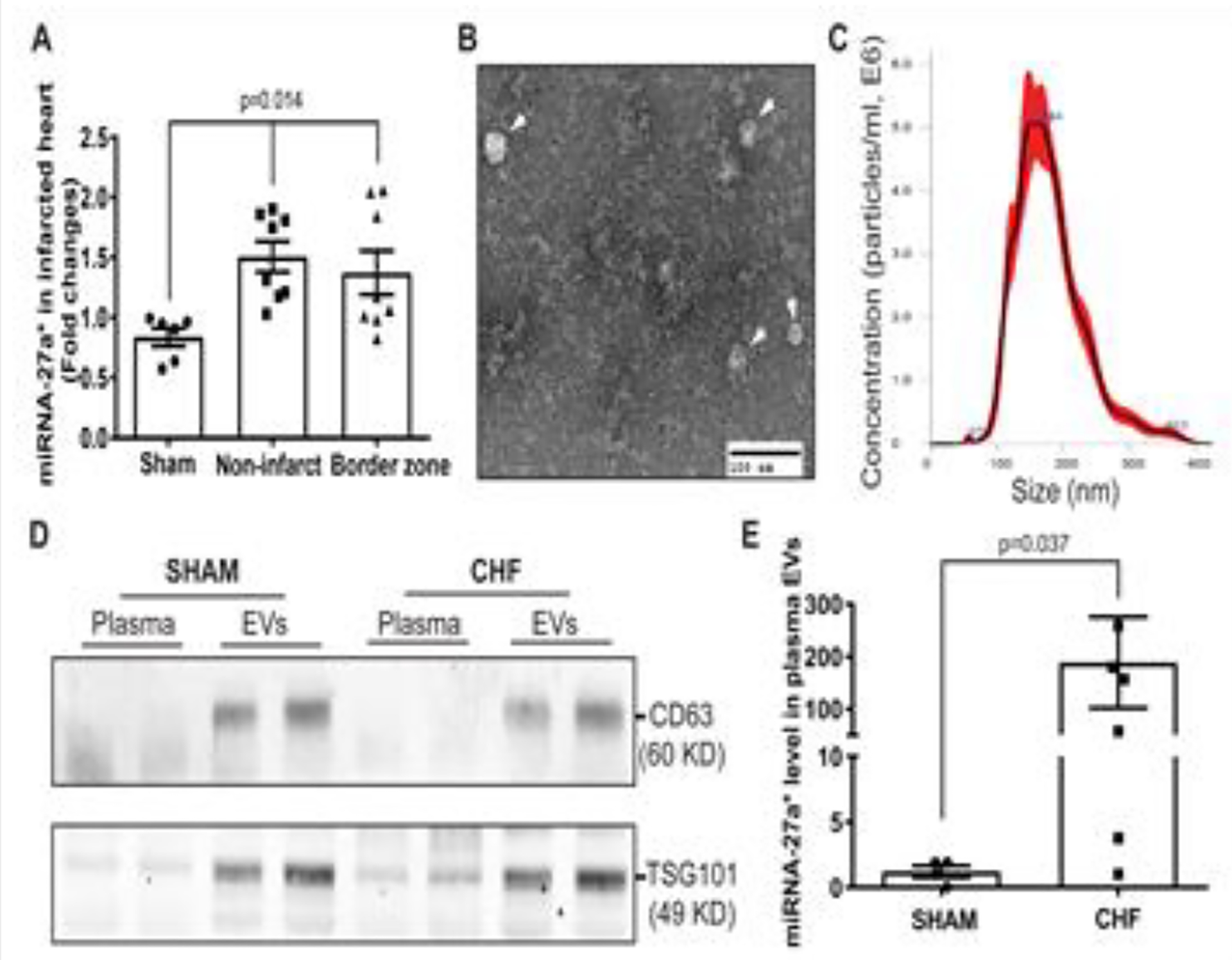
Figure 1. miRNA-27a* is up-regulated in the infarcted heart and circulating extracellular vesicles.

qRT-PCR shows that miRNA-27a* is up-regulated in the both non-infarcted and border zone tissues of the left ventricle compared to that in Sham rats (A), U6 snRNA was used as an internal control (±SEM, n=6, Sham; n=8, CHF); EVs isolated from rat plasma by differential centrifugation were subjected to negative staining and electron microscopy (B). Scale bar is 100 nm; EVs were also subjected to Malvern NanoSight analysis show the size distribution of circulating EVs. Five captures were collected and averaged (±SEM, n=5) (C); Western blotting data shows the typical EV markers (CD63 and TSG101) in EV pellets derived from plasma of Sham and CHF rats, respectively (D); qRT-PCR results showing increased miRNA-27a* in plasma EVs from CHF rats compared to that of Sham rats (E), cel-mir-39 was used as a spike-in control (±SEM, n=6).
