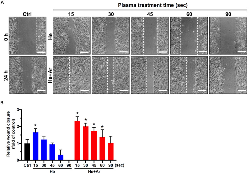
FIGURE 3.

CAPJ facilitates human keratinocyte migration. HaCaT cells were treated with PAM that was pretreated with He-CAPJ or He/Ar-CAPJ for the indicated times. The cells were then subjected to a wound healing assay and cultured for 24 h to analyze cell migration activity. (A) The images were acquired after scratching for 0 or 24 h. Scale bars, 300 μm. (B) Cell migration activity was assessed by determining the relative wound closure, as described in section “Materials and Methods.” The data were presented as the mean ± standard deviation of three independent experiments. Statistical significance was determined using the Student’s t-test. *P < 0.05 when compared to the control group.
