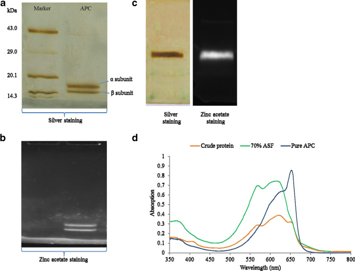
Fig. 1.
APC purification and characterization by PAGE and UV–visible spectrum analysis. a Purified APC isolated from Phormidium sp. A09DM was analyzed on 15% SDS-PAGE. Protein standard molecular weight markers are shown in the lane denoted as “Marker” and two distinct bands of α (15.5 kDa) and β (17.7 kDa) subunits were observed on the SDS-PAGE stained with silver nitrate. b Zinc acetate staining of 15% SDS-PAGE confirms the presence of two distinct fluorescence subunits α (15.5 kDa) and β (17.7 kDa). c Native PAGE showed the intact purified APC stained with silver nitrate and zinc acetate. d Overlay UV–visible spectrum of crude, 70% ASF and pure APC. Λmax at 653 nm confirmed the presence of APC. Increase in the peak at 653 nm was observed after concurrent purification steps

