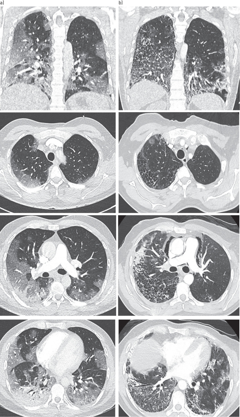
FIGURE 1.
a) Initial CT scan: images show extensive ground-glass opacities in both lungs with subpleural predominance affecting 50–75% of lung parenchyma, as well as condensation in the right lower lobe. b) Control CT imaging at day-10 : axial CT images show extensive honeycombing cysts associated with septal thickening, with subpleural predominance (where ground-glass opacities had initially been observed, notably in the right lower and upper lobes), with associated traction bronchiectasis. Pneumomediastinum also developed. There was no sign of pulmonary embolism.

