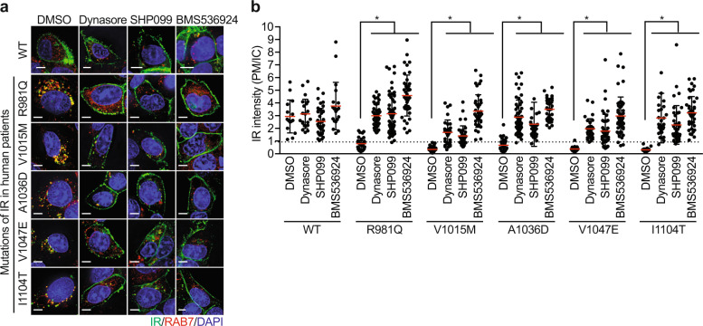
Fig. 4. Characterization of class II IR mutations found in human patients.
a HepG2 cells stably expressing IR-GFP WT or Class II mutants were serum starved for 14 h, treated with the indicated inhibitors for 4 h, and stained with anti-GFP (IR; green) and anti-RAB7 (Red; D95F2, Cell Signaling) antibodies. Dynasore (dynamin inhibitor, 80 μM, Sigma), SHP099 (SHP2 inhibitor, 10 μM, MedChemExpress), and BMS536924 (IR kinase inhibitor, 2 μM, Tocris). Scale bar, 5 μm. b Quantification of the ratios of PM and IC IR-GFP signals of the cells shown in a (mean ± SD; *p < 0.0001).

