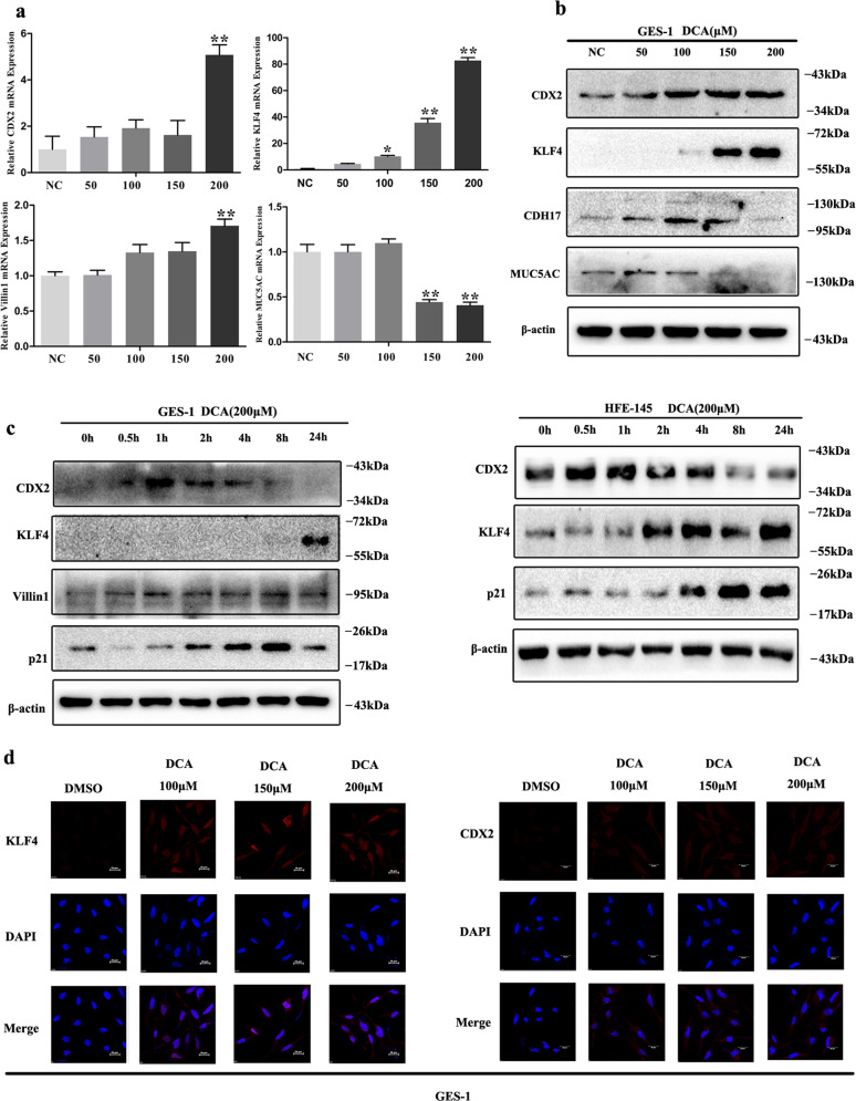
Fig. 1. Deoxycholic acid (DCA) treatment induced metaplasia markers expression in gastric epithelial cells.
a, b GES-1 cells were treated with different doses of DCA for 24 h. Next, CDX2, KLF4, Villin1, and MUC5AC mRNA were detected by qRT-PCR. Error bar indicates the SEM, *P < 0.05, **P < 0.01 vs. negative control (NC), n = 3. CDX2, KLF4, CDH17, and MUC5AC proteins were detected by western blotting (WB). c GES-1 (upper) and HFE-145 (lower) cells were treated with 200 μM DCA in a time-dependent manner. Columnar genes (CDX2, KLF4, Villin1, and p21) were examined by WB. d GES-1 cells were treated with 200 μM DCA for 24 h. KLF4 (left) and CDX2 (right) expression was analyzed by immunofluorescent staining (red). Nucleus was stained with DAPI (blue). Scale bar, 20 μm.

