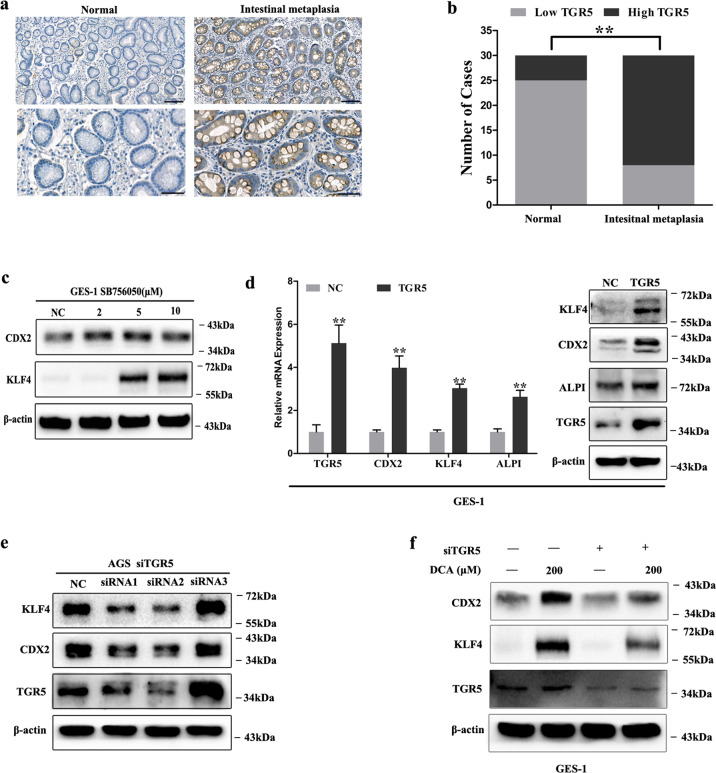
Fig. 2. TGR5 was involved in gastric intestinal metaplasia (IM) development.
a Immunohistochemical staining of normal and IM tissues showing TGR5 expression. Scale bar, 100 μm (upper) and 50 μm (lower). b Column charts showed the cases with high and low TGR5 expression in IM tissues. **P < 0.01. c GES-1 cells were treated with SB756050 (2, 5, and 10 μM) for 24 h. Then, KLF4 and CDX2 protein expression was examined by WB. d GES-1 cells were transfected with TGR5 overexpression lentiviral. TGR5, KLF4, CDX2, and ALPI mRNA and protein levels were analyzed by qRT-PCR and WB. Error bar indicates the SEM, **P < 0.01 vs. NC, n = 3. e AGS cells were transfected with siRNAs target TGR5 for 72 h. Then TGR5, KLF4, and CDX2 protein expressions were examined by WB. f Further, TGR5 was knocked down by transfection with siRNA in GES-1 cells. Next, the cells were treated with DCA (200 μM) for 24 h. TGR5, KLF4, and CDX2 were examined by WB.

