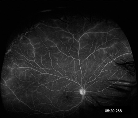
Figure 15.

Fluorescein angiography with ultra-wide-field imaging shows vascular leakage in the superior and temporal periphery in addition to the optic disc and macular leakage. Shadowing caused by the lashes is present in the inferior and nasal regions
