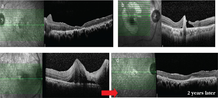
Figure 16.

Macular atrophy in a patient with advanced Behçet’s uveitis (a), macular atrophy and hole in another patient (b), a patient who presented with active retinitis involving the macula and associated macular edema (c), and the same patient 2 years later, exhibiting disorganization and atrophy of the retinal layers and subfoveal fibrosis (d)
