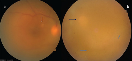
Figure 2.

Diffuse vitritis and vitreous haze are observed in two different Behçet’s uveitis patients. In the first patient (a), the optic disc is hyperemic and there is a small retinitis focus (white arrow) at the posterior pole. In the other patient (b), the vitreous haze is very dense and the optic disc (black arrow) and ghost vessels below (blue arrows) are barely discernible
