Figure 7.

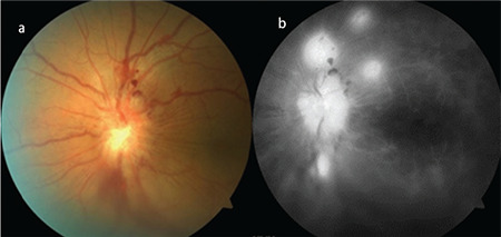
Color fundus photographs (a) and fluorescein angiography images (b) of a Behçet’s patient who developed optic disc neovascularization. There is extensive vascular and capillary leakage

Color fundus photographs (a) and fluorescein angiography images (b) of a Behçet’s patient who developed optic disc neovascularization. There is extensive vascular and capillary leakage