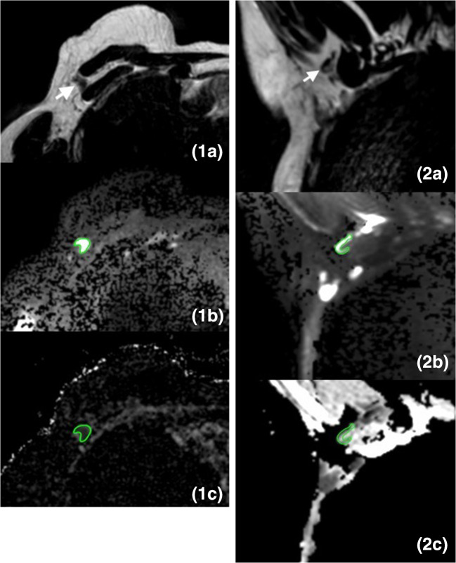
Fig. 3.
Images of a 27-mm large invasive ductal carcinoma in the right breast of a 64-year-old female patient, which was treated with mastectomy and ALND (pT2N1). The white arrow points to an axillary lymph node. The green delineation shows an axillary lymph node. 1a Axial T2W breast MR image shows the axillary lymph node with the longest axis. 1b Axial DW breast MR image (b-value = 800 s/mm2) shows the same axillary lymph node with relatively high signal intensity. 1c Axial ADC map of breast MRI shows corresponding lymph node with relatively low signal intensity with an ADC value of 0.646 × 10⁻³ mm2/s. 2a Coronal T2W dedicated axillary MR image shows the axillary lymph node with the longest axis. 2b Coronal DW dedicated axillary MR image (b-value = 800 s/mm2) shows the same lymph node with relatively high signal intensity. 2c Coronal ADC map of dedicated axillary MRI shows corresponding lymph node with relatively low signal intensity with an ADC value of 0.837 × 10⁻³ mm2/s

