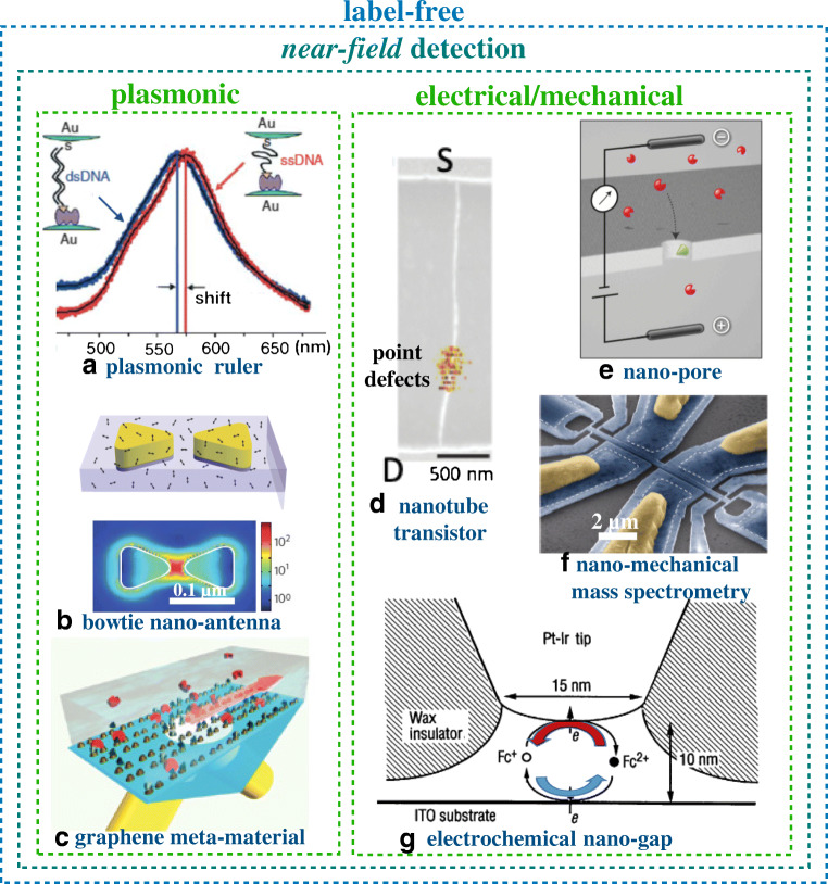
Fig. 2.
Single-molecule assays with near-field detections. a Spectral shift of the plasmonic signal generated by a pair of gold particles connected by a single-stranded DNA (red curve) and a double-stranded DNA (blue curve) (Reprinted with permission from Ref. [9] Copyright 2005 Springer Nature). b Schematic structure of a gold bow tie nanoantenna in a thin poly(methyl methacrylate) film doped with a near-infrared N,N′-bis(2,6-diisopropylphenyl)-1,6,11,16-tetra-[4-(1,1,3,3-tetramethylbutyl)phenoxy]quaterrylene-3,4:13,14-bis(dicarboximide) (TPQDI) dye (top panel) and the result of the modelling of local intensity enhancement in the bow tie structure (bottom panel) (Reprinted with permission from Ref. [10] Copyright 2009 Springer Nature). c Schematics of the biosensing apparatus entailing a plasmonic metamaterial. Biotin (recognition element) is represented by dark green triangles and streptavidin (analyte) by red disks. Light beams are shown in yellow while the grazing diffracted wave is represented by a red arrow (Reprinted with permission from Ref. [11] Copyright 2013 Springer Nature). d The single carbon nanotube transistor is shown along with the source (S) and drain (D) pads. The gating is performed through a phosphate-buffered saline solution at pH 7.4. The electrochemically generated point defects serve as anchoring sites to covalently bind single-stranded DNA probes (Reprinted with permission from Ref. [12] Copyright 2011 Springer Nature). e Cross-sectional sketch featuring a nanopore system through which charged single molecules translocate. Charged analyte molecules are depicted in red while the recognition element is colored in green (Reprinted with permission from Ref. [13] Copyright 2012 Springer Nature). f Electron micrograph of a multimode nano-electromechanical based mass detection. Yellow regions represent Al/Si gate contacts. Narrow gauges near the ends of the fingers become strained with the motion of the finger, thereby transducing mechanical motion into electrical resistance (Reprinted with permission from Ref. [14] Copyright 2012 Springer Nature). g Schematic illustration of the tip–substrate configuration in electrochemical nanowells engaging one electroactive species (Reprinted with permission from Ref. [15] Copyright 1995 Science)

