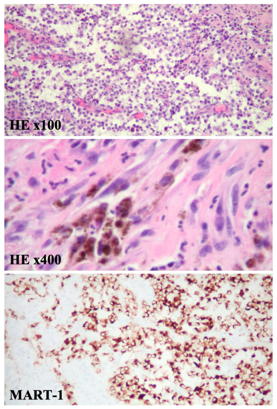
Figure 3. Pathology demonstrates a malignant melanoma.

At medium power magnification, histopathology reveals the tumor cells are round with hyperchromatic eccentric nuclei, prominent nucleoli, and mixed eosinophilic and basophilic cytoplasm. Some areas reveal necrosis with neutrophilic exudate. Mitotic activity is frequently seen (HE x100). Pleomorphic melanoma cells containing various amounts of brown melanin pigment (HE x400). At high power magnification, tissue stained with MART-1 (melan-A) by IHC show melanocytes highlighted in brown.
