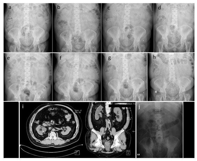
Figure 3. Sequential imaging photos.
Imaging after right ureterorenoscopy, right nephrostomy, and right percutaneous nephrolithotomy in April 2016 ( a), imaging after ureterorenoscopy and percutaneous nephrolithotomy in June 2016 ( b), imaging after the second extracorporeal shock wave lithotripsy in June 2016 ( c), imaging after the third extracorporeal shock wave lithotripsy in July 2016 ( d), imaging after right laser ureterorenoscopy and replacement of right double J stent in July 2016 ( e), imaging after extracorporeal shock wave lithotripsy in October 2016 ( f), imaging after double J stent removal in October 2016 ( g), imaging as a routine control in January 2018 ( h & i), imaging after retrograde intrarenal surgery which shows no residual stone in June 2019 ( j).

